Comprehensive Cardiac Diagnostic Services
At A Glance
Quick Menu
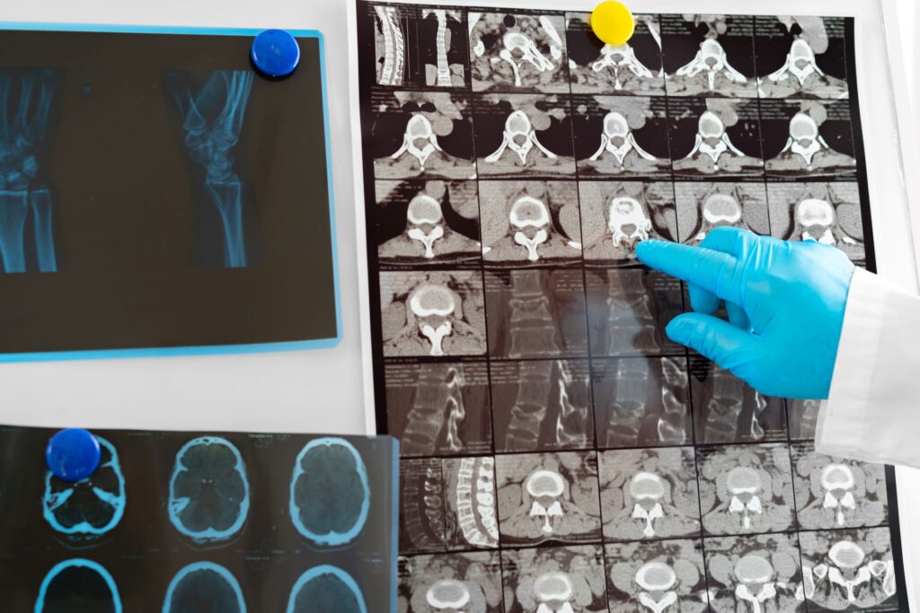
Accurate diagnosis is the foundation of effective cardiac care. At Cardiac Care Specialists of Dublin, we offer a complete range of diagnostic services performed right in our office using state-of-the-art technology.
All testing is conducted by our experienced clinical team, and Dr. Malhotra personally interprets every study. This hands-on approach ensures diagnostic accuracy and allows us to provide results typically within 24 hours.
Whether you’re experiencing symptoms or need routine monitoring of an existing condition, our diagnostic services provide the detailed information necessary to guide your treatment plan.
Nuclear Stress Testing (C-Cam Technology)
Nuclear stress testing is one of the most accurate methods for evaluating blood flow to your heart muscle. Using our advanced C-Cam nuclear imaging system, this test creates detailed pictures of your heart during rest and physical stress, revealing areas with reduced blood flow that may indicate coronary artery disease.
The test is particularly valuable for detecting “silent” heart disease in patients without obvious symptoms, evaluating chest pain to determine if it’s heart-related, assessing the severity of known coronary artery disease, and checking how well previous treatments like bypass surgery or stent placement are working.
Dr. Malhotra is board-certified in nuclear cardiology and personally interprets all nuclear stress tests performed at our practice. This expertise ensures accurate diagnosis and appropriate treatment recommendations based on your test results.

What to Expect During the Test
A nuclear stress test typically takes 3 to 4 hours and involves several steps:
Resting Images:
You'll receive a small injection of a radioactive tracer (the amount of radiation is minimal and safe). After waiting 30-60 minutes for the tracer to circulate, we'll take initial images of your heart at rest using our C-Cam nuclear camera.
Stress Phase:
You'll exercise on a treadmill or stationary bike while we monitor your heart rate, blood pressure, and EKG. If you're unable to exercise, we can use medication to simulate the effects of exercise on your heart.
Stress Images:
Dr. Malhotra will compare the resting and stress images to identify any areas of reduced blood flow or heart muscle damage.
Results:
Dr. Malhotra will compare the resting and stress images to identify any areas of reduced blood flow or heart muscle damage.
- Resting Images: You'll receive a small injection of a radioactive tracer (the amount of radiation is minimal and safe). After waiting 30-60 minutes for the tracer to circulate, we'll take initial images of your heart at rest using our C-Cam nuclear camera.
- Stress Phase: You'll exercise on a treadmill or stationary bike while we monitor your heart rate, blood pressure, and EKG. If you're unable to exercise, we can use medication to simulate the effects of exercise on your heart.
- Stress Images: After reaching your target heart rate, you'll receive a second tracer injection. We'll then take another set of images showing blood flow to your heart during stress.
- Results: Dr. Malhotra will compare the resting and stress images to identify any areas of reduced blood flow or heart muscle damage.
Who Needs a Nuclear Stress Test?
Your doctor may recommend nuclear stress testing if you:
- Experience chest pain, shortness of breath, or other symptoms that might indicate heart disease
- Have risk factors for coronary artery disease (diabetes, high blood pressure, high cholesterol, family history, smoking)
- Need evaluation before surgery
- Have had previous heart problems and require ongoing monitoring
- Have abnormal results from other cardiac tests
- Are being evaluated for treatment effectiveness after bypass surgery, stent placement, or heart attack
Benefits of Nuclear Stress Testing
Nuclear stress testing offers several important advantages:
- Early Detection: Identifies coronary artery disease before symptoms become severe, allowing earlier intervention and better outcomes.
- Comprehensive Assessment: Provides detailed information about blood flow patterns, heart function, and the extent of any blockages.
- Treatment Guidance: Helps Dr. Malhotra determine whether you need medication adjustments, lifestyle changes, or procedures like angioplasty or bypass surgery.
- Risk Stratification: Assesses your risk for future heart problems, helping prioritize preventive measures.
- Non-Invasive: Unlike cardiac catheterization, nuclear stress testing doesn't require inserting catheters into your blood vessels.
- Repeatable: Can be performed multiple times to track changes over time without cumulative radiation concerns.
Early Detection:
Identifies coronary artery disease before symptoms become severe, allowing earlier intervention and better outcomes.
Comprehensive Assessment:
Provides detailed information about blood flow patterns, heart function, and the extent of any blockages.
Treatment Guidance:
Helps Dr. Malhotra determine whether you need medication adjustments, lifestyle changes, or procedures like angioplasty or bypass surgery.
Risk Stratification:
Assesses your risk for future heart problems, helping prioritize preventive measures.
Non-Invasive:
Unlike cardiac catheterization, nuclear stress testing doesn't require inserting catheters into your blood vessels.
Repeatable:
Can be performed multiple times to track changes over time without cumulative radiation concerns.
Preparing for Your Test
To ensure accurate results:
- Avoid caffeine for 24 hours before your test (no coffee, tea, soda, chocolate, or energy drinks)
- Don't eat or drink anything except water for 4 hours before your appointment
- Wear comfortable clothing and walking shoes
- Bring a list of all your medications
- Tell us if you have asthma or use an inhaler
- Inform us if you're pregnant or might be pregnant
If you take certain heart medications, Dr. Malhotra may ask you to temporarily stop them before the test. Our staff will provide specific instructions when you schedule your appointment.
Echocardiograms (GE Vivid IQ System)
An echocardiogram uses sound waves to create live, moving images of your heart as it beats. This completely non-invasive test provides detailed information about your heart’s structure, valve function, and pumping efficiency.
We use the GE Vivid IQ echocardiography system, which represents the latest advancement in cardiac ultrasound technology. This portable, high-resolution system delivers exceptionally clear images that help Dr. Malhotra make accurate diagnoses.
Echocardiography is valuable for diagnosing many cardiac conditions including heart valve disease, heart failure, cardiomyopathy, congenital heart defects, and fluid around the heart (pericardial effusion). It also measures your ejection fraction, an important indicator of how well your heart pumps blood with each beat.
The test is completely painless and safe for patients of all ages.
Types of Echocardiograms We Perform
Transthoracic Echocardiogram (TTE):
This is the standard echocardiogram where a technician places a transducer (ultrasound wand) on your chest. Gel is applied to improve sound wave transmission. The technician moves the transducer to different positions to capture images of your heart from multiple angles. The test typically takes 30 to 60 minutes.
Stress Echocardiogram:
This test combines echocardiography with exercise or medication to evaluate how your heart performs under stress. We take ultrasound images before and immediately after exercise to identify problems that may not be apparent when your heart is at rest.
What an Echocardiogram Reveals
Your echocardiogram provides crucial information about:
- Heart Size and Shape: Identifies enlarged chambers or abnormally thick heart walls that might indicate various cardiac conditions.
- Pumping Strength: Measures ejection fraction, showing what percentage of blood your heart pumps out with each beat. Normal is 55-70%.
- Valve Function: Shows how well your heart valves open and close. Detects stenosis (narrowing) or regurgitation (leaking).
- Blood Flow: Uses Doppler imaging to visualize blood flow patterns and detect abnormal flow.
- Heart Muscle Motion: Identifies areas of the heart muscle that aren't contracting properly, which might indicate prior heart attack or reduced blood supply.
- Fluid Around the Heart: Detects pericardial effusion that could affect heart function.
Heart Size and Shape:
Identifies enlarged chambers or abnormally thick heart walls that might indicate various cardiac conditions.
Pumping Strength:
Measures ejection fraction, showing what percentage of blood your heart pumps out with each beat. Normal is 55-70%.
Valve Function:
Shows how well your heart valves open and close. Detects stenosis (narrowing) or regurgitation (leaking).
Blood Flow:
Uses Doppler imaging to visualize blood flow patterns and detect abnormal flow.
Heart Muscle Motion:
Identifies areas of the heart muscle that aren't contracting properly, which might indicate prior heart attack or reduced blood supply.
Fluid Around the Heart:
Detects pericardial effusion that could affect heart function.
Benefits of Echocardiography
- Completely non-invasive with no needles, radiation, or contrast dye
- Painless and requires no recovery time
- Safe for all ages, including pregnant women
- Provides real-time visualization of heart function
- Can be repeated as often as necessary without risk
- Relatively quick test (30-60 minutes)
- Immediately identifies many cardiac problems
- Helps guide treatment decisions
Preparing for Your Echocardiogram
- Wear comfortable, loose-fitting clothing
- You may eat and drink normally before the test
- Take your regular medications unless instructed otherwise
- Bring a list of your current medications
- Arrive 10 minutes early to complete paperwork
For stress echocardiograms, additional preparation similar to stress testing may be required. Our staff will provide specific instructions.
Extended Cardiac Monitoring (30-Day)
Many heart rhythm problems are intermittent, occurring unpredictably and sometimes without noticeable symptoms. A standard EKG performed during an office visit captures only a brief snapshot of your heart rhythm. Extended cardiac monitoring provides continuous or event-based recording over 30 days, dramatically increasing the likelihood of detecting arrhythmias.
We use advanced monitoring devices from Zoll and Phillips, industry leaders in cardiac monitoring technology. These lightweight, discreet devices allow you to go about your normal daily activities while they continuously track your heart rhythm.
When you experience symptoms like palpitations, dizziness, shortness of breath, or chest discomfort, you can mark the event. The monitor records your heart’s electrical activity during these episodes, allowing Dr. Malhotra to correlate your symptoms with actual heart rhythm data.

Types of Cardiac Monitors
Holter Monitor (24-48 Hours):
A traditional Holter monitor provides continuous recording for one to two days. You wear small electrodes attached to a portable recording device. This shorter monitoring period is useful when symptoms occur frequently.
Extended Holter Monitor (7-14 Days):
Similar to standard Holter monitoring but worn for up to two weeks, increasing the chance of capturing infrequent arrhythmias.
Event Monitor (30 Days):
Event monitors record your heart rhythm continuously but store data only when you activate the device during symptoms or when the device automatically detects an abnormal rhythm. This 30-day monitoring period is ideal for capturing sporadic episodes.
Mobile Cardiac Telemetry:
These advanced monitors continuously transmit data in real-time, allowing immediate review if dangerous arrhythmias occur. They provide the most comprehensive monitoring available.
Who Needs Cardiac Monitoring?
Extended cardiac monitoring is recommended if you experience:
- Palpitations or sensation of skipped heartbeats
- Unexplained dizziness or lightheadedness
- Fainting spells (syncope)
- Shortness of breath without clear cause
- Chest discomfort that comes and goes
- Fatigue that might be rhythm-related
- Previous stroke with suspected cardiac cause
- Known arrhythmia requiring monitoring
Monitoring is also used to check how well arrhythmia medications are working or to evaluate pacemaker function.
What Cardiac Monitoring Detects
- Extended monitoring can detect:
- Atrial Fibrillation (AFib): Irregular, often rapid heart rhythm in the upper chambers that increases stroke risk.
- Atrial Flutter: Similar to AFib but with a more regular pattern.
- Supraventricular Tachycardia (SVT): Episodes of rapid heartbeat originating above the ventricles.
- Ventricular Tachycardia: Potentially dangerous rapid rhythm from the lower heart chambers.
- Bradycardia: Abnormally slow heart rate that may cause symptoms.
- Heart Block: Delays or interruptions in electrical signal transmission.
- Premature Beats: Extra heartbeats that may or may not require treatment depending on frequency and symptoms.
Atrial Fibrillation (AFib):
Irregular, often rapid heart rhythm in the upper chambers that increases stroke risk.
Atrial Flutter:
Similar to AFib but with a more regular pattern.
Supraventricular Tachycardia (SVT):
Episodes of rapid heartbeat originating above the ventricles.
Ventricular Tachycardia:
Potentially dangerous rapid rhythm from the lower heart chambers.
Bradycardia:
Abnormally slow heart rate that may cause symptoms.
Heart Block:
Delays or interruptions in electrical signal transmission.
Premature Beats:
Extra heartbeats that may or may not require treatment depending on frequency and symptoms.
Benefits of 30-Day Monitoring
- Increased Detection Rate: The longer monitoring period significantly increases the likelihood of capturing intermittent arrhythmias. Studies show 30-day monitoring detects up to 3 times more arrhythmias than 24-hour Holter monitors.
- Symptom Correlation: Allows precise matching of your symptoms to heart rhythm at that exact moment, definitively determining if symptoms are heart-related.
- Treatment Optimization: Provides comprehensive data to guide medication adjustments or decisions about procedures.
- Peace of Mind: If monitoring shows normal rhythms during your symptoms, it reassures you that your heart isn't the problem.
- Stroke Prevention: Early detection of atrial fibrillation allows prompt initiation of blood thinners to prevent stroke.
What to Expect
- Modern cardiac monitors are surprisingly easy to use:
- Small adhesive electrodes attach to your chest
- The recording device is compact and lightweight
- Most devices are water-resistant for showering
- You can sleep, work, and perform most normal activities
- Keep a symptom diary noting when you feel palpitations, dizziness, or other symptoms
- Press the event button if your device has one when symptoms occur
- The device stores data for Dr. Malhotra to review
- Our staff provides thorough instructions on using your specific monitor type.
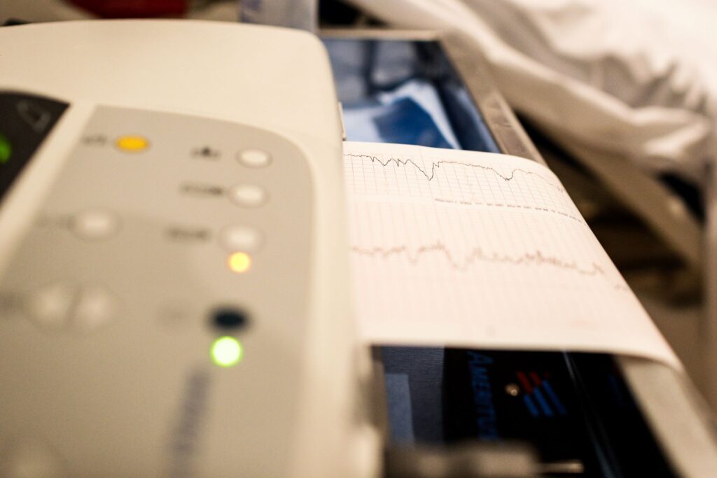
Electrocardiogram (EKG/ECG)
An electrocardiogram (EKG or ECG) records your heart’s electrical activity. This quick, painless test provides valuable information about your heart rate, rhythm, and electrical conduction patterns.
EKGs are performed during most cardiology visits and are essential for detecting many cardiac conditions. The test takes only a few minutes and involves placing small electrodes on your chest, arms, and legs. These electrodes detect the electrical signals that control your heartbeat.
While a single EKG provides a snapshot of your heart’s electrical activity, it’s often combined with other tests for comprehensive evaluation.
What an EKG Can Detect
An EKG helps diagnose:
- Heart rhythm abnormalities (arrhythmias)
- Heart attack (current or previous)
- Inadequate blood flow to the heart muscle (ischemia)
- Enlarged heart chambers
- Electrolyte imbalances
- Medication effects on the heart
- Pacemaker function
- Congenital heart defects
When You Need an EKG
EKGs are performed:
- During routine cardiology evaluations
- When you experience chest pain, palpitations, or shortness of breath
- Before certain surgeries
- To monitor heart medication effects
- During stress testing
- To follow up on abnormal previous EKGs
- As part of annual physical exams for patients at cardiac risk
The test is completely safe, non-invasive, and has no risks or side effects.
Fast Results, Expert Interpretation
One of the key advantages of having all testing performed in our office is rapid result turnaround. Dr. Malhotra personally interprets every diagnostic study, ensuring consistency and accuracy in your care.
Most test results are available within 24 hours. In urgent situations, Dr. Malhotra can provide preliminary findings even faster. Once your results are ready, we’ll contact you to discuss the findings and any recommended next steps.
If additional testing or treatment is needed, we’ll explain why and schedule follow-up appointments promptly. You’ll never be left wondering what your results mean or what to do next.
Understanding Your Results
We believe you should fully understand your test results. Dr. Malhotra or one of our nurse practitioners will:
- Explain what the test showed in clear, understandable terms
- Discuss what the findings mean for your heart health
- Answer all your questions
- Recommend next steps if treatment or additional testing is needed
- Provide written copies of your results for your records
- Send reports to your referring physician if applicable
Schedule Your Test Today
Scheduling diagnostic testing is easy. Call our office at (478) 246-2264, and our staff will:
- Verify your insurance coverage and benefits
- Find an appointment time that fits your schedule
- Provide preparation instructions for your specific test
- Answer questions about what to expect
- Send appointment reminders
Most tests can be scheduled within a few days. If you’re experiencing urgent symptoms, we’ll prioritize your appointment appropriately.